

Table of Contents
Pregnancy Screening in Vizag | Fetal Scans & Genetic Diagnostics
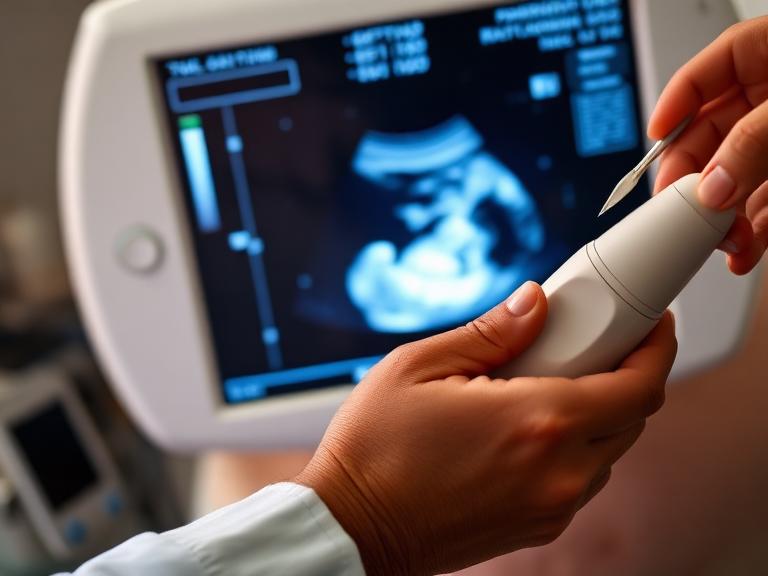
Pregnancy screening forms the core of modern maternal-fetal medicine. Systematic prenatal screening allows maternal health specialists to monitor embryology, verify gestational age, evaluate placental perfusion, and identify structural or chromosomal abnormalities at the earliest possible stage. Seeking expert Pregnancy Screening in Vizag ensures access to advanced ultrasonography, maternal serum biomarker analysis, and non-invasive prenatal testing (NIPT) within structured clinical settings.
Across Visakhapatnam—from medical centers in Health City (Arilova), MVP Colony, and Maharani Peta to Siripuram, Jagadamba Junction, and Sheela Nagar—fetal medicine facilities integrate high-resolution sonography with clinical genetics. These diagnostic procedures provide expectant parents and obstetricians with actionable clinical insights throughout every trimester.
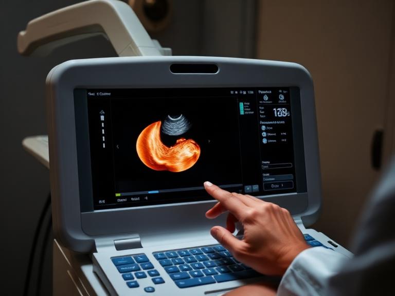
Clinical Protocol: Trimester-Wise Screening Pathway
Diagnostic evaluations follow specific gestational windows to track both maternal physiological adaptations and fetal organogenesis.
                         [Trimester-Wise Screening Pathway]
                                         │
     ┌───────────────────────────────────┼───────────────────────────────────┐
     ▼                                   ▼                                   ▼
[First Trimester]               [Second Trimester]                  [Third Trimester]
(Weeks 6–13+6)                  (Weeks 18–22)                       (Weeks 28–40)
 • Viability & Dating Scan       • Level II Anomaly Scan             • Fetal Growth & Wellbeing Scan
 • Nuchal Translucency (NT)      • Fetal Echocardiography            • Umbilical & Uterine Doppler
 • First Trimester Combined      • Maternal Serum Quad Screen        • Biophysical Profile (BPP)
 • NIPT / cell-free DNA          • Cervical Length (Transvaginal)    • Gestational Diabetes (75g OGTT)
Core Diagnostics in Pregnancy Screening
1. First Trimester Diagnostics (Weeks 6 to 13+6)
- Early Viability & Dating Ultrasound (Weeks 6–9): Establishes crown-rump length (CRL), verifies intra-uterine implantation site, rules out ectopic pregnancy, and checks cardiac reactivity.
- Nuchal Translucency (NT) Scan (Weeks 11–13+6): Measures the fluid-filled space behind the fetal neck. An increased NT measurement (>3.0 mm) can indicate higher risks for trisomies (Down syndrome, Edwards syndrome, Patau syndrome) or structural cardiac anomalies.
- First Trimester Combined Screening (FTCS): Integrates NT thickness measurements with double marker biochemical blood tests—specifically Free Beta-hCG and PAPP-A (Pregnancy-Associated Plasma Protein A)—to estimate risk probabilities.
- Non-Invasive Prenatal Testing (NIPT): Analyzes cell-free fetal DNA (cfDNA) circulating in maternal blood to detect aneuploidies with over 99% sensitivity.
2. Second Trimester Diagnostics (Weeks 18 to 22)
- Detailed Anomaly Scan (Level II / TIFFA Scan): Evaluates overall fetal anatomy, including intracranial structures, facial profile, four-chamber heart view, abdominal wall, renal pelves, spine, and long bones.
- Fetal Echocardiography (Weeks 18–24): Focused specialized ultrasound for high-risk pregnancies (e.g., maternal diabetes, family history of congenital heart defects) to rule out complex cardiovascular malformations.
- Quadruple Marker Serum Screen (Weeks 15–20): Evaluates Maternal Serum Alpha-Fetoprotein (MSAFP), Total hCG, Unconjugated Estriol ($uE3$), and Inhibin-A when first-trimester screening was missed.
- Transvaginal Cervical Length Audit: Measures internal cervical os length to screen for cervical insufficiency and assess preterm birth risks.
3. Third Trimester Diagnostics (Weeks 28 to 40)
- Fetal Growth & Wellbeing Scan: Tracks estimated fetal weight (EFW), abdominal circumference (AC), head circumference (HC), and amniotic fluid index (AFI) to detect Intrauterine Growth Restriction (IUGR) or fetal macrosomia.
- Color Doppler Velocimetry: Assesses blood flow dynamics within the umbilical artery, middle cerebral artery (MCA), and maternal uterine arteries to check placental sufficiency.
- Biophysical Profile (BPP): Combines Non-Stress Testing (NST) with ultrasound monitoring of fetal tone, body movements, breathing, and amniotic fluid volume.
- 75g Oral Glucose Tolerance Test (OGTT): Routinely administered between 24 and 28 weeks to screen for Gestational Diabetes Mellitus (GDM).
Trimester Diagnostic Matrix
| Gestational Window | Clinical Test / Procedure | Diagnostic Focus & Clinical Purpose |
| Weeks 6 – 9 | Viability & Dating Scan | Confirms intrauterine sac, cardiac activity, gestational age, and chorionicity in twin pregnancies. |
| Weeks 11 – 13+6 | NT Scan + Double Marker Test | Evaluates nuchal fold thickness and serum PAPP-A/Free Beta-hCG to screen for Down syndrome. |
| Weeks 10+ Onward | NIPT (cfDNA Blood Test) | Directly sequences cell-free fetal DNA in maternal blood for chromosomal trisomies (21, 18, 13) and sex chromosome aneuploidies. |
| Weeks 18 – 22 | Anomaly (TIFFA / Level II) Scan | High-resolution evaluation of all internal organ systems, face, spine, limbs, and placental attachment. |
| Weeks 18 – 24 | Fetal Echocardiogram | Targeted cardiovascular ultrasound to inspect cardiac chambers, valves, and major outflow tracts. |
| Weeks 24 – 28 | 75g OGTT (Glucose Screening) | Identifies maternal glucose intolerance and gestational diabetes mellitus. |
| Weeks 28 – 36 | Fetal Growth & Doppler Study | Tracks growth curves, amniotic fluid levels (AFI), and umbilical/cerebral vascular resistance. |
| Weeks 36 – 40 | Biophysical Profile (BPP) & NST | Evaluates fetal oxygenation, acute neuro-central reflexes, and placental reserve prior to labor. |
Screening Progression Sequence
1.1. Pregnancy Confirmation & Dating Scan:Weeks 6–9 Gestation.
Confirm intrauterine implantation, visualize fetal pole, record embryonic heart rate, and establish precise gestational dating.
2.2. First Trimester Chromosomal Risk Audit:Weeks 11–13+6 Gestation.
Perform the NT scan alongside Double Marker serum testing (or NIPT) to assess early chromosomal risks and confirm structural development.
3.3. Targeted Anomaly (TIFFA) Scan:Weeks 18–22 Gestation.
Conduct a detailed structural anatomical scan to evaluate internal organ development, facial structures, neural tube integrity, and placental positioning.
4.4. Maternal Metabolic Screening:Weeks 24–28 Gestation.
Administer a 75g Oral Glucose Tolerance Test (OGTT) to assess maternal glycemic health and rule out Gestational Diabetes Mellitus.
5.5. Fetal Growth & Hemodynamic Doppler Assessment:Weeks 28–36 Gestation.
Monitor fetal weight gain, evaluate amniotic fluid levels, and check vascular resistance using umbilical artery Doppler imaging.
Key Screening Centers & Fetal Medicine Units in Vizag
Visakhapatnam offers access to advanced maternal-fetal diagnostics across several major medical hubs:
- Arilova (Health City): Tertiary multi-specialty hospitals with specialized fetal medicine departments capable of advanced imaging, high-risk pregnancy monitoring, and invasive diagnostics.
- Maharani Peta & Jagadamba Junction: Home to established diagnostic scan centers and mother-and-child hospitals with maternal-fetal imaging infrastructure.
- MVP Colony & Siripuram: Clinical diagnostic hubs offering high-resolution 3D/4D ultrasound, first-trimester screening, and fetal medicine consultations.
- Sheela Nagar & Gajuwaka: Healthcare facilities providing routine prenatal care, anomaly scans, and maternal health monitoring for suburban residential belts.

Frequently Asked Questions (FAQs)
1. What is the primary purpose of pregnancy screening in Vizag?
Pregnancy screening identifies structural, genetic, or growth-related abnormalities in the fetus, alongside maternal conditions like preeclampsia and gestational diabetes. Early screening allows timely management and planning throughout prenatal care.
2. How is a screening test different from a diagnostic test?
Screening tests (e.g., Double Marker, NT scan, NIPT) calculate the risk probability of a condition. Diagnostic tests (e.g., Chorionic Villus Sampling or Amniocentesis) provide definitive confirmation when a screening test returns a high-risk result.
3. What is an NT scan, and when is it performed?
The Nuchal Translucency (NT) scan is an ultrasound conducted between 11 weeks and 13 weeks + 6 days. It measures fluid accumulation at the back of the baby’s neck to assess Down syndrome and cardiac risk factors.
4. What is the Level II / TIFFA scan, and why is it crucial?
The Target Scan for Fetal Anomalies (TIFFA) occurs between 18 and 22 weeks. It evaluates fetal anatomy—including the brain, heart, spine, stomach, kidneys, and limbs—to identify structural malformations.
5. What is Non-Invasive Prenatal Testing (NIPT)?
NIPT is a maternal blood test that analyzes cell-free fetal DNA (cfDNA) circulating in maternal blood. It screens for chromosomal trisomies (21, 18, and 13) with high accuracy (>99%) starting from week 10 of pregnancy.
6. What blood tests are included in the Double Marker test?
The Double Marker test measures two maternal serum biomarkers: Free Beta-hCG and PAPP-A (Pregnancy-Associated Plasma Protein A). Combining these levels with the NT scan measurement improves screening accuracy for trisomies.
7. Why is a Doppler ultrasound performed during the third trimester?
Doppler ultrasounds evaluate blood circulation in the umbilical artery, middle cerebral artery, and uterine arteries. This assesses placental performance, ensures the fetus receives adequate oxygen, and helps detect intrauterine growth restriction (IUGR).
8. How is Gestational Diabetes Mellitus (GDM) screened?
GDM screening uses a 75g Oral Glucose Tolerance Test (OGTT), typically between 24 and 28 weeks. Fasting, 1-hour, and 2-hour blood glucose levels are evaluated after drinking a standardized glucose solution.
9. What is a Fetal Echocardiogram?
A Fetal Echocardiogram is a targeted ultrasound examination of the fetal heart conducted by a fetal medicine specialist, typically between 18 and 24 weeks. It checks for congenital heart defects in high-risk pregnancies.
10. Are ultrasound scans safe for the baby during pregnancy?
Yes. Medical diagnostic ultrasounds use sound waves rather than ionizing radiation. They have been used for decades and are safe for both mother and baby when performed by qualified imaging professionals.
11. What is the Quadruple Marker test?
The Quadruple Marker test is a second-trimester maternal blood screening test performed between 15 and 20 weeks. It measures AFP, Total hCG, Unconjugated Estriol ($uE3$), and Inhibin-A to estimate risk for trisomies and neural tube defects.
12. How is cervical length evaluated during pregnancy screening?
Cervical length is evaluated using transvaginal ultrasound, often alongside the Level II anomaly scan. Shortened cervical length (<25 mm) indicates a higher risk of preterm labor, allowing preventive steps like cervical cerclage or progesterone therapy.
13. What is a Biophysical Profile (BPP)?
A Biophysical Profile evaluates fetal health during the third trimester. It combines Non-Stress Test (NST) electronic heart rate monitoring with ultrasound assessments of fetal breathing, body movement, muscle tone, and amniotic fluid volume.
14. What should I do if a screening test indicates a high risk?
A high-risk result does not mean a definitive diagnosis. Your fetal medicine specialist will offer genetic counseling and discuss diagnostic options, such as NIPT or amniocentesis, to confirm the findings.
15. What are the typical costs for pregnancy screening tests in Vizag?
- First Trimester NT Scan + Double Marker: ₹2,500 – ₹5,000
- Detailed Anomaly Scan (TIFFA / Level II): ₹2,000 – ₹4,500
- Non-Invasive Prenatal Testing (NIPT): ₹12,000 – ₹25,000
- Third Trimester Growth Scan with Doppler: ₹2,000 – ₹4,000